Вопрос:
This is a multiple-choice question related to biology. Identify the correct characteristic of the tissue shown in the image.
Ответ:
Analysis of the Image:
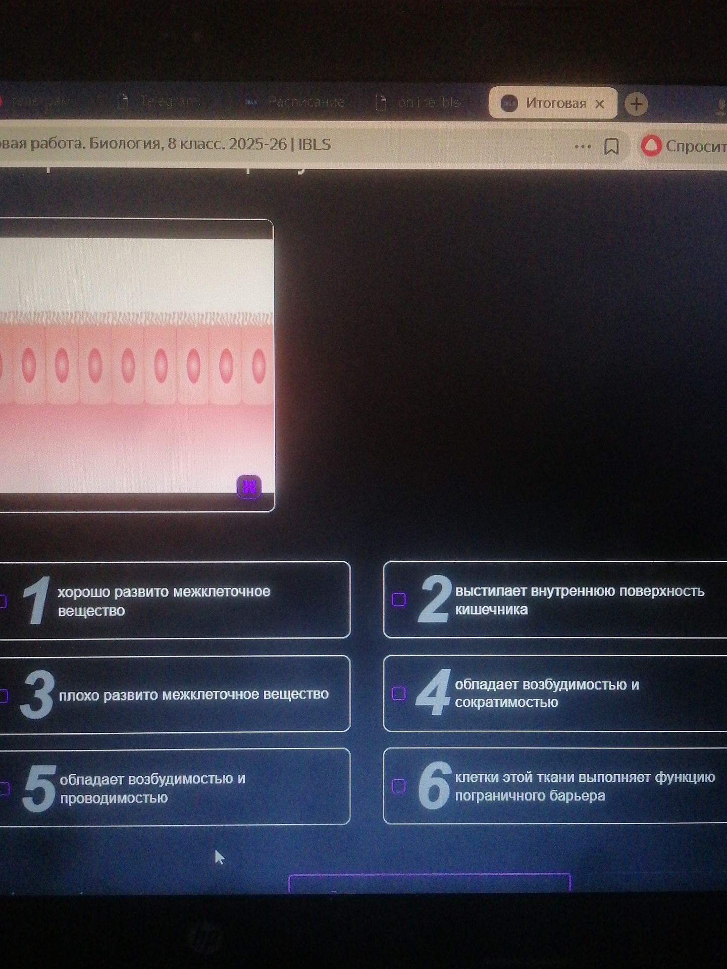
The image displays a magnified view of a single layer of epithelial cells, characterized by their columnar shape and a brush border (microvilli) on the apical surface. The presence of microvilli significantly increases the surface area for absorption. This type of epithelial tissue is commonly found lining the small intestine and other absorptive surfaces.
Let's analyze the options provided:
- 1. хорошо развито межклеточное вещество (well-developed intercellular substance): Epithelial tissues generally have very little intercellular substance, as their cells are tightly packed. Connective tissues have abundant intercellular substance.
- 2. выстилает внутреннюю поверхность кишечника (lines the inner surface of the intestine): This is a characteristic function and location of the type of tissue shown, especially with the presence of microvilli for absorption.
- 3. плохо развито межклеточное вещество (poorly developed intercellular substance): While true for epithelial tissue, this is a general characteristic and not the most specific or functionally descriptive option in this context.
- 4. обладает возбудимостью и сократимостью (possesses excitability and contractility): These are characteristics of muscle tissue, not epithelial tissue.
- 5. обладает возбудимостью и проводимостью (possesses excitability and conductivity): These are characteristics of nervous tissue, not epithelial tissue.
- 6. клетки этой ткани выполняет функцию пограничного барьера (cells of this tissue perform the function of a boundary barrier): While epithelial tissues do form a barrier, option 2 is more specific to the visual evidence of absorptive surface enhancement (microvilli).
Based on the visual evidence (columnar cells with microvilli) and the typical functions of such tissues, option 2 is the most accurate description.
Ответ: 2. выстилает внутреннюю поверхность кишечника
